Your Vision, Our Care: Premier Eye Care Services in Boise

Eye care that goes beyond the routine

Intermountain Eye Centers’ primary eye care provides an entry point for patients to receive vision tests and glasses or contact lenses, screenings for eye diseases, diagnosis and treatment of most eye conditions. If needed, our doctors will refer patients to specialists and coordinate with other aspects of medical care.

Primary eye care services

  1. Educating patients about maintaining and promoting healthy vision.
  2. Performing a comprehensive examination of the visual system.
  3. Screening for eye diseases and conditions affecting vision.
  4. Recognizing ocular manifestations of systemic diseases and systemic effects of ocular medications.
  5. Making diagnoses for any detected abnormalities.
  6. Performing refractions.
  7. Fitting and prescribing optical aids such as glasses and contact lenses.
  8. Counseling and educating patients about their eye disease conditions.
  9. Recognizing and managing the effects of drug therapy.
  10. Performing surgery when necessary.

When are eye examinations necessary?

  • Providers recommend an eye exam every two years for normal findings, and more often if managing or treating eye-care symptoms.
  • Persons with diabetes are at risk for several eye disorders and should have eye examinations every year.
  • Patients 65 and older should typically have an eye examination annually.
  • African-Americans are four to five times more likely to develop glaucoma than Caucasians.
  • Our optometrists see patients 6 years and older. Younger patients are referred to a pediatric specialist in the Treasure Valley.

We treat and screen for several disorders

During an eye exam, an eye doctor reviews your medical history and completes a series of tests to determine the health of your eyes. The information from an eye exam may lead to medical procedures or prescriptions.

Frequently Asked Questions

What is dry eye?

Dry eye is one of the most common problems treated by eye physicians. It can be caused by several factors, including the quality of the tear film that lubricates the eye.

Detection and treatment

Symptoms of dry eye include redness, burning, excessive tearing, blurred vision, light sensitivity and foreign body sensations. One of the most common causes of dry eye is the normal aging process, but environmental factors, contact lens use, certain medical conditions and medications may also contribute. Dry eyes are more pronounced in persons who have reduced blink frequency, such as persons who frequently use the computer. Patients may also have increased discomfort after periods of reading, driving or watching TV. The physicians at Intermountain Eye Centers are experienced and committed to the treatment of dry eyes and related disorders. We take a multifaceted approach and address tear production, quality, distribution, drainage and maintenance. Careful management by our eye-care professionals can significantly improve a patient’s symptoms and quality of life.

Facts about contact lenses

About 36 million Americans wear contact lenses, 80 percent of whom wear daily wear soft lenses. Currently, there are four types of contact lenses in use:

  • Soft, water-absorbing lenses
  • Rigid, gas-permeable lenses

Reading a contact lens prescription

The prescription for contact lenses includes more information than what is available on the prescription for eyeglasses. Special measurements will need to be taken of the curvature of the eye. In addition, the physician will determine if the eyes are too dry for contact lenses and if there are any corneal problems that may prevent a person from wearing contact lenses. Trial lenses are usually tested on the eyes for a period of time to ensure proper fit.

The contact lens prescription usually includes the following information:

  • Contact lens power (measured in diopters, like eyeglasses)
  • Contact lens base curve
  • Diameter of the lens
  • Type of material

Eye-care specialists are required by federal law to give you a copy of your contact lens specifications.

When scratches create further problems

The cornea is the clear front window that covers the colored portion of the eye. A corneal erosion or abrasion refers to a scratch or injury to the outer layer of the cornea. Common causes of these painful abrasions include contact lenses, a baby’s fingernail, tree limbs, bushes or vigorous rubbing of the eye.

The most common treatment for corneal erosions is to patch the eye tightly. An antibiotic may be prescribed to prevent infection. A corneal erosion usually heals within a week.

If bacteria get into the tissues under the protective layer of the eye a corneal ulcer can occur. These complications can be very serious and cause loss of vision. Proper care by you and your doctor is necessary to prevent serious consequences.

The return of chickenpox

A patient’s first encounter with the herpes zoster virus is usually childhood chickenpox. Later in life, the virus may reactivate, causing a rash of small blisters, frequently on the chest or forehead. This second encounter is known as shingles.

It is important to see a doctor when herpes zoster occurs on the face because the virus may invade the eye. A doctor will look for elevated pressure in the eye, inflammation and herpes zoster lesions on the surface of the eye. These problems can be treated but they are sometimes difficult to manage, so careful treatment and follow-up are required. New oral antivirus medications can provide patients with a quicker, more complete recovery.

Fighting the elements

A pterygium is a fibrous, fleshy growth on the surface of the clear cornea. It occurs most frequently in people who are exposed to lots of sun, wind, dust or harsh climates. Pterygium is most commonly seen in the tropics and areas with wide temperature swings, but it is also seen in temperate climates among people who work or spend much of their time outdoors. Pterygium is three times more common in men than women.

Sometimes patients mistake pterygium for cataracts, but cataracts are not as easily seen with the naked eye as pterygium is.

The best form of therapy is prevention, such as wearing hats and dark glasses in bright sunshine. If clear vision is threatened by the presence of a pterygium a surgical incision may be necessary.

Normal vision vs. Cataract vision, which is much blurrier

Types of cataracts

According to the National Eye Institute, cataract types are subdivided accordingly:

  • Age-related cataracts: The majority of cataracts are related to aging.
  • Congenital cataracts: Some babies are born with cataracts or develop them in childhood, often in both eyes. Some congenital cataracts do not affect vision, but others do and need to be removed.
  • Secondary cataracts: Secondary cataracts develop primarily as a result of another disease occurrence in the body (i.e., diabetes). Secondary cataract development has also been linked to steroid use.
  • Traumatic cataracts: Eyes that have sustained an injury may develop traumatic cataracts either immediately after the incident, or several years later.

Other sources, including the American Academy of Ophthalmology, describe the different types of cataracts according to the cataract location on the eye lens, including:

  • Nuclear cataract: This is the most common type of cataract and the most common type associated with aging. Nuclear cataracts develop in the center of the lens and can induce nearsightedness, a temporary improvement in reading vision which is sometimes referred to as “second sight.” Unfortunately, “second sight” disappears as the cataract grows.
  • Cortical cataract: This type of cataract initially develops as wedge-shaped spokes in the cortex of the lens, with the spokes extending from the outside of the lens to the center. When these spokes reach the center of the lens they interfere with the transmission of light and cause glare and loss of contrast. This type of cataract is frequently developed in persons with diabetes, and while it usually develops slowly, it may impair both distance and near vision so significantly that surgery is often suggested at an early stage.
  • Subcapsular cataract: A subcapsular cataract usually starts as a small opacity under the capsule, at the back of the lens. This type of cataract develops slowly and significant symptoms may not occur until the cataract is well developed. A subcapsular cataract is often found in persons with diabetes, myopia, retinitis pigmentosa, and in those taking steroids.

What is Conjunctivitis?

Conjunctivitis, also known as “pink eye,” is an inflammation of the conjunctiva of the eye. The conjunctiva is the membrane that lines the inside of the eye and also a thin membrane that covers the actual eye.

What causes conjunctivitis?

There are many different causes of conjunctivitis. The following are the most common causes:

  • Bacteria, including:
    • Staphylococcus aureus
    • Haemophilus influenza
    • Streptococcus pneumoniae
    • Neisseria gonorrhea
    • Chlamydia trachomatis
  • Viruses, including:
    • adenoviruses
    • herpes virus
  • Chemicals (seen mostly in the newborn period after the use of medicine in the eye to prevent other problems)
  • Allergies

What are the different types of conjunctivitis?

Conjunctivitis is usually divided into at least two categories: newborn conjunctivitis and childhood conjunctivitis. There are different causes and treatments for each.

NEWBORN CONJUNCTIVITIS

The following are the most common causes and treatment options of newborn conjunctivitis:

  • Chemical conjunctivitis: This is related to an irritation in the eye from the use of eye drops that are given to the newborn to help prevent a bacterial infection. Sometimes, the newborn reacts to the drops and may develop a chemical conjunctivitis. The eyes are usually mildly red and inflamed, starting a few hours after the drops have been placed in the eye, and lasts for only 24 to 36 hours. This type of conjunctivitis usually requires no treatment.
  • Gonococcal conjunctivitis: This is caused by a bacteria called Neisseria gonorrhea. The newborn obtains this type of conjunctivitis by the passage through the birth canal from an infected mother. This type of conjunctivitis may be prevented with the use of eye drops in newborns at birth. The newborn eyes usually are very red, with thick drainage and swelling of the eyelids. This type usually starts about two to four days after birth. Treatment for gonococcal conjunctivitis usually will include antibiotics through an intravenous (IV) catheter.
  • Inclusion conjunctivitis: This is caused by an infection with chlamydia trachomatis, obtained by passage through the birth canal from an infected mother. The symptoms include moderate thick drainage from the eyes, redness of the eyes, swelling of the conjunctiva, and some swelling of the eyelids. This type of conjunctivitis usually starts 5 to 12 days after birth. Treatment usually will include oral antibiotics.
  • Other bacterial causes: After the first week of life, other bacteria may be the cause of conjunctivitis in the newborn. The eyes may be red and swollen with some drainage. Treatment depends on the type of bacteria that has caused the infection. Treatment usually will include antibiotic drops or ointments to the eye, warm compresses to the eye, and proper hygiene when touching the infected eyes.

CHILDHOOD CONJUNCTIVITIS

Childhood conjunctivitis is a swelling of the conjunctiva and may also include an infection. It is a very common problem in children. Also, large outbreaks of conjunctivitis are often seen in daycare settings or schools. The following are the most common causes of childhood conjunctivitis:

  • Bacteria
  • Viral
  • Allergies
  • Herpes

What are the symptoms of childhood conjunctivitis?

The following are the most common symptoms of childhood conjunctivitis. However, each child may experience symptoms differently. Symptoms may include:

  • Itchy, irritated eyes
  • Clear, thin drainage (usually seen with viral or allergic causes)
  • Sneezing and runny nose (usually see with allergic causes)
  • Stringy discharge from the eyes (usually seen with allergic causes)
  • Thick, green drainage (usually seen with bacterial causes)
  • Ear infection (usually seen with bacterial causes)
  • Lesion with a crusty appearance (usually seen with herpes infection)
  • Eyes that are matted together in the morning
  • Swelling of the eyelids
  • Redness of the conjunctiva
  • Discomfort when the child looks at a light
  • Burning in the eyes

The symptoms of conjunctivitis may resemble other medical conditions or problems. Always consult your child’s doctor for a diagnosis.

How is conjunctivitis diagnosed?

Conjunctivitis is usually diagnosed based on a complete medical history and physical examination of your child’s eye. Cultures of the eye drainage are usually not required, but may be done to help confirm the cause of the infection.

Treatment for conjunctivitis

Specific treatment for conjunctivitis will be determined by your physician based on:

  • Your child’s age, overall health, and medical history
  • Extent of the condition
  • Your child’s tolerance for specific medications, procedures, or therapies
  • Expectations for the course of the condition
  • Your opinion or preference

Specific treatment depends on the underlying cause of the conjunctivitis.

  • Bacterial causes: Your child’s physician may order antibiotic drops to put in the eyes.
  • Viral causes: Viral conjunctivitis usually does not require treatment. Your child’s physician may order antibiotic drops for the eyes to help decrease the chance of a secondary infection.
  • Allergic causes: Treatment for conjunctivitis caused by allergies usually will involve treating the allergies. Your child’s physician may order oral medications or eye drops to help with the allergies.
  • Herpes: If your child has an infection of the eye caused by a herpes infection, your child’s physician may refer you to an eye care specialist. Your child may be given both oral medications and eye drops. This is a more serious type of infection and may result in scarring of the eye and loss of vision.

Infection can be spread from one eye to the other, or to other people, by touching the affected eye or drainage from the eye. Proper hand washing is very important. Drainage from the eye is contagious for 24 to 48 hours after beginning treatment.

Normal vision vs. Glaucoma vision, which is characterized by darkness at the edges of vision

What is glaucoma?

Glaucoma is a condition in which the normal fluid pressure inside the eyes (intraocular pressure, or IOP) slowly rises as a result of the fluid aqueous humor – which normally flows in and out of the eye – not being able to drain properly. Instead, the fluid collects and causes pressure damage to the optic nerve (a bundle of more than 1 million nerve fibers that connects the retina with the brain) and loss of vision.

What causes glaucoma?

While physicians used to think that high intraocular pressure (also known as ocular hypertension) was the main cause of optic nerve damage in glaucoma, it is now known that even persons with normal IOP can experience vision loss from glaucoma. Thus, the causes are still unknown.

Diabetes and its effects on eyes

Diabetic eye disease comprises a group of eye conditions that affect people with diabetes. These conditions include diabetic retinopathy, diabetic macular edema (DME), cataract, and glaucoma. All forms of diabetic eye disease have the potential to cause severe vision loss and blindness.

Diabetic eye disease is a group of eye conditions that can affect people with diabetes. Diabetic retinopathy affects blood vessels in the light-sensitive tissue called the retina that lines the back of the eye. It is the most common cause of vision loss among people with diabetes and the leading cause of vision impairment and blindness among working-age adults. Diabetic macular edema (DME) is a consequence of diabetic retinopathy. It is swelling in an area of the retina called the macula. Diabetic eye disease also includes cataract and glaucoma.

The early stages of diabetic retinopathy usually have no symptoms. The disease often progresses unnoticed until it affects vision. Bleeding from abnormal retinal blood vessels can cause the appearance of “floating” spots. These spots sometimes clear on their own. But without prompt treatment, bleeding often recurs, increasing the risk of permanent vision loss. If DME occurs, it can cause blurred vision.

Diabetic retinopathy and DME can be detected during a comprehensive dilated eye exam.

What is age-related macular degeneration?

Age-related macular degeneration (AMD) is a disease that affects an individual’s central vision. AMD is the most common cause of visual impairment in patients over the age of 55 in industrialized nations. It produces variable symptoms, ranging from no symptoms at all to profound central visual loss. Because only the center of vision is affected, people rarely go blind from this disease. However, AMD can make it difficult of read, drive, or perform other daily activities that require fine, central vision.

AMD occurs when the macula, which is located in the center of the retina and provides us with sight in the center of our field of vision, begins to degenerate. With less of the macula working, central vision – which is necessary for driving, reading, recognizing faces, and performing close-up work – begins to deteriorate.

What are the different types of AMD?

There are two primary types of AMD:

  • Dry Form: This type of AMD is the most common. The atrophic or dry form of AMD is marked by the presence of drusen, pigmentary, or atrophic changes in the center of the retina, i.e. the macula. At this stage, symptoms are typically mild, although some vision loss may occur. Although the dry form is currently untreatable, certain patients may benefit from prophylaxis with vitamins and antioxidants, although such use must be carefully supervised to prevent unwanted side-effects.
  • Wet Form: This type of AMD is less common, but accounts for almost all severe vision loss caused by either type of AMD. A minority but very important group of AMD patients will develop the wet form, marked by the growth of new blood vessels under the macula (choroidal neovascularization) with associated bleeding and leakage with central vision loss. The wet form of AMD requires urgent attention and expert care, because it is treatable in many cases with the intraocular injection of anti-VEGF (vascular endothelial growth factor) medications such as Lucentis, Avastin, and others in development. Although the injections themselves are easily tolerated and not painful, the decisions regarding initiation of treatment and ongoing maintenance are complex and require care in the evaluation of diagnostic tests such as Fluorescein Angiography and Optical Coherence Tomography (OCT). AMD, while clearly a continuing problem of epidemic proportions, is increasingly a treatable condition as a result of many successful laboratory and clinical investigations.

A lump on the surface of the eye

A pinguecula is a small, yellowish lump on the surface of the white part of the eye. It appears at 3 o’clock and 9’oclock, if the eye is viewed as a clock. Dryness and exposure to the environment usually cause these fleshy elevations. People who work or spend much of the time outdoors are most prone to this condition.

Symptoms may include burning or stinging of the eyes. Sometimes a pinguecula may become red, irritated and inflamed. Treatment with appropriate drops will typically clear this redness in just a few days. In rare cases, surgery is necessary.

Scary, but usually a cosmetic problem

A subconjunctival hemorrhage appears as a sharply outlined, bright red spot on the white part of the eye. Most of the time, no symptoms accompany these hemorrhages. Many people become alarmed by the sudden onset of this common problem, even though it is not associated with any diseases that cause vision loss.

The most common cause of a subconjunctival hemorrhage is simple rubbing of the eyes. Only rarely is the condition associated with high blood pressure or other bleeding problems. No treatment is needed. Subconjunctival hemorrhages will go away and only be a cosmetic problem for a few days.


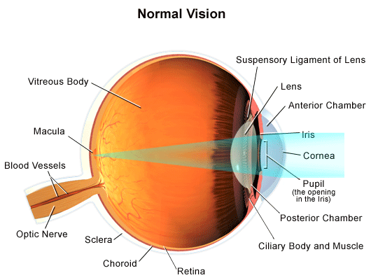
How normal vision happens

In order to better understand how refractive errors affect our vision, it is important to understand how normal vision occurs. For persons with normal vision, the following sequence takes place:

  1. Light enters the eye through the cornea, the clear, dome-shaped surface that covers the front of the eye.
  2. From the cornea, the light passes through the pupil. The amount of light passing through is regulated by the iris, or the colored part of your eye.
  3. From there, the light then hits the lens, the transparent structure inside the eye that focuses light rays onto the retina.
  4. Next, it passes through the vitreous humor, the clear, jelly-like substance that fills the center of the eye and helps to keep the eye round in shape.
  5. Finally, it reaches the retina, the light-sensitive nerve layer that lines the back of the eye, where the image appears inverted.
  6. The optic nerve is then responsible for interpreting the impulses it receives into images.

Diagram of light passing through the eye

What are refractive errors?

The following are the most common refractive errors, all of which affect vision and may require corrective lenses or surgery for correction or improvement.

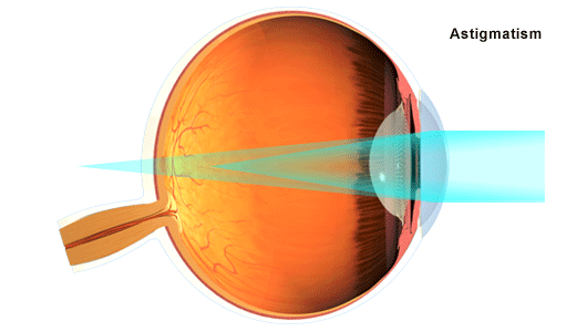
ASTIGMATISM

astigmatism
Astigmatism is a condition in which an abnormal curvature of the cornea can cause two focal points to fall in two different locations – making objects up close and at a distance appear blurry. Astigmatisms may cause eye strain and may be combined with nearsightedness or farsightedness. Eyeglasses, contact lenses, or corrective surgery may help to correct or improve the condition.

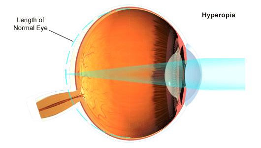
HYPEROPIA
hyperopia

Commonly known as farsightedness, hyperopia is the most common refractive error in which an image of a distant object becomes focused behind the retina, either because the eyeball axis is too short, or because the refractive power of the eye is too weak. This condition makes close objects appear out of focus and may cause headaches and/or eye strain.

Eyeglasses or contact lenses may help to correct or improve hyperopia by adjusting the focusing power to the retina. Corrective surgery may also help by changing the shape of the cornea to a more spherical, round shape instead of an oval shape.

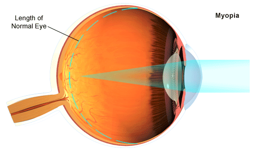
MYOPIA

myopia

Commonly known as nearsightedness, myopia is a condition in which, opposite of hyperopia, an image of a distant object becomes focused in front the retina, either because the eyeball axis is too long, or because the refractive power of the eye is too strong. This condition makes distant objects appear out of focus and may cause headaches and/or eye strain.

Eyeglasses or contact lenses may help to correct or improve myopia by adjusting the focusing power to the retina. Corrective surgery may also help by changing the shape of the cornea to a more spherical, round shape instead of an oblong shape.

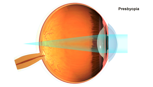
PRESBYOPIA

presbyopia
Another type of farsightedness, presbyopia is caused when the center of the eye lens hardens making it unable to accommodate near vision. This condition generally affects almost everyone over the age of 50 – even those with myopia. Eyeglasses or contact lenses may be prescribed to correct or improve the condition.

“ After consulting with a few other eye surgeons in the area I was impressed with both the technology at Intermountain Eye Care and the caring and professionalism of the staff. I've been very satisfied with my care before, during, and after my procedure. My LASIK surgery went flawlessly, my recovery went smoothly, and my vision is perfect. Without reservation I recommend Dr. Fethke and Intermountain Eye Center. ”